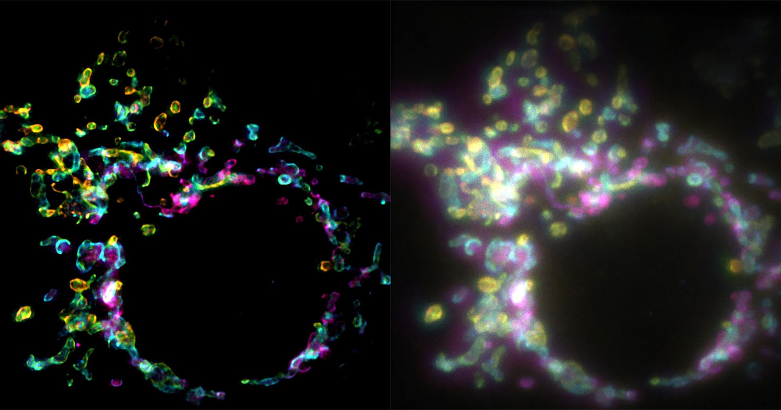
Super-resolution microscopy has one big weakness when it comes to living things: it is slow. Most widefield techniques build a single sharp image out of hundreds or even thousands of frames, and cells do not sit still while that happens. A team at EPFL in Switzerland has found a way around the problem by making use of a property of light that ordinary imaging ignores. Their method is called SPIFFI, and it produces a super-resolved picture from one camera exposure.
SPIFFI stands for spatial polarization-induced fluorescence fluctuation imaging. It was developed by Wei Guo, Lely Feletti and Aleksandra Radenovic at the Laboratory of Nanoscale Biology (LBEN) in EPFL’s School of Engineering. The paper, titled “SPIFFI enables single-shot super-resolution and multidimensional imaging”, was published in Nature Methods, and EPFL presented the work on September 8, 2026. An earlier version was posted as a preprint on bioRxiv on December 15, 2025.
Polarization instead of time
Fluorescent molecules do not glow evenly in every direction. The light they emit is polarized, with its waves oscillating preferentially in certain directions depending on how each molecule is oriented. SPIFFI splits the fluorescence into four polarization-sensitive channels and compares the resulting images to pull out structural detail that a standard widefield microscope blurs together.
“Essentially, previous approaches used temporal information to resolve spatial resolution, but this doesn’t work very well on living cells,” says first author and LBEN PhD student Wei Guo. SPIFFI takes the information from the polarization channels of a single frame instead of from a long stack of frames.
What it can resolve
- Resolution improvement of up to twofold from a single snapshot
- Structures around 160–170 nanometers in size resolved in one exposure
- About 80 nanometers when SPIFFI images are post-processed with existing fluctuation-based methods
- Four polarization-sensitive detection channels
- Demonstrated on both fixed and live cells, including mitochondrial and microtubule dynamics and events such as cellular splitting and fusion
Speed is the real gain. “With previous techniques, taking many images would only result in one super-resolved frame. With SPIFFI, every frame is super-resolved, meaning we can now produce super-resolution videos of live cells,” Guo explains. Radenovic says the method “can capture fast-moving processes within cells, while enabling high-throughput, multi-dimensional imaging beyond the limits of conventional microscopes.”
The comparison images released by EPFL make the point clearly. Next to a conventional widefield image, the mitochondrial outer membrane captured with SPIFFI shows much cleaner outlines instead of a soft glow.
Beyond sharper pictures
Because the setup measures polarization directly, it records more than intensity. The preprint’s abstract describes SPIFFI as a multi-channel polarimetric method capable of six-dimensional information extraction. The authors also say the single-shot approach makes volumetric live-cell super-resolution imaging much more feasible.
Just as important for real labs, SPIFFI’s optical hardware can be integrated into existing fluorescence microscopes, so it does not require a dedicated instrument. The researchers are working on a more compact version of the setup and on combining the technique with three-dimensional imaging, with live-cell biology, neuroscience, biophysics and drug discovery named as possible uses.
For anyone who thinks about imaging in terms of sensors and optics, SPIFFI is a neat reminder that resolution is not only about pixels and lenses. Sometimes the extra detail is already in the light, waiting for the right way to read it.